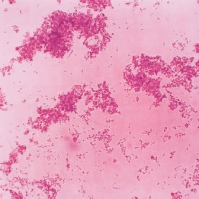
Encephalomyocarditis virus: Difference between revisions
imported>Daniel Mietchen (formatting) |
imported>Daniel Mietchen m (Encephalomyocarditis Virus moved to Encephalomyocarditis virus: naming conventions) |
Revision as of 01:55, 23 April 2009
For the course duration, the article is closed to outside editing. Of course you can always leave comments on the discussion page. The anticipated date of course completion is May 21, 2009. One month after that date at the latest, this notice shall be removed. Besides, many other Citizendium articles welcome your collaboration! |
 |
| Scientific classification |
|
|
Description and Significance
Encephalomyocarditis Virus(EMCV) is a member of the genus Cardiovirus of the family Picornaviridae. It's said that the Pacornavirusinfects many animal species, including pigs, rodents, cattle, elephants, raccons, marsupials, and primates such as baboons, monkeys, chimpanzees, as well as humans. There are two type of Encephalomyocarditis Virus. One is Encephalomyocarditis Virus type A, which causes reproductive problems. The other one is Encephalomyocarditis Virus type B, which causes heart failure in pigs. African Elephants were the first species that were infected with the virus. The first outbreak ever seen was in South Africa in 1993. Between December 1993 and August 1994, a number of acute deaths occurred in free-ranging Africa elephants in the Kruger National Park KNP. 4
Genome and Structure
The main host of Encephalomyocarditis Virus(EMCV) are the rat and mouse. The virus is passed through fecal oral transmission. This discovery was documented after a large population explosion in rodents during the same time as a large of number of elephants where dying. Encephalomyocarditis Virus(EMCV) attacks many animals as I have mention int he beginning but as I read articles the one that has been documented and studied the most has been the pig. This is virus causes acute myocarditis and sudden death in preweaned pigs, whereas trans placental infections of sows cause fetal mummification, abortion, still birth,and neonatal death. 4 Once humans are infected with this virus, the symptoms they may be faced with include fever, neck stiffness, lethargy, delirium, headaches, and vomiting. In primates such as gibbons and owl monkeys, Encephalomyocarditis Virus can cause necrotizing and interstitial myocarditis.[6] Transmission and pathogenesis occurs by ; incubation from nine to ten days, oral, fecal and urine contamination of food, sub clinical infections, replication in myocardial and kills them.
Cell Structure and metabolism
Some diagnoses may be antermorten due to rapid clinical course, gross lesions such as pale streak in the myocardium, hydrothorax, hydropericardium, pulmonary edema, froth in tracheobronchial tree. Other diagnoses are Histopathology :myocardial degeneration and necrosis with lymphocytic infiltrates, virus particles may be visible on electron microscopy, definitive diagnosis: virus isolation, PCR, mouse inoculation, serological test for antibodies available-Texas A&m. 5
Ecology and Pathology
The only treatment I have read about actually a treatment, but prevention, the oil-adjuvant Encephalomyocarditis vaccine. This vaccine has been given to elephants, mice and pigs so far. Also, scientist are finding that controlling the rodent population is crucial to preventing the spread of this disease. Immune prophylaxis is considered to be another one of the effective strategies for controlling this virus in pigs and other animals who may possibly carry the virus. In humans it's very rare to get this virus.
Application to Biotechnology
There are many methods used to futher study the effects of this virus. One was the viruse isolation and serology, which consisted on inoculation of a infected tissue of the bull elephant in the Kruger National Park (KNP)into mice. Vaccine seed virus was prepared by adapting this E1-M1 isolated by passing five time on a monolayer of BHK 21 clone 13 cells which are used at the laboratory for routine foot-and-mouth disease (FMD) vaccine production. 4 Seology is the virus neutralization test that is used as antigen. Another way is the oil adjuvant which is the double suspension which was used as vaccine for pigs, elephants, and mice. Virus titrtion is flat-bottomed micro titer plates using an in-house modification and tryptose, referred to as Vac medium, for the dilution of the virus and suspension of the BHK cells. The plates where incubated at 37 Celsius for forty-eight hours in humidified chamber containing five percent of carbon dioxide and the test read with an inverted microscope. The last one mentioned in the journal was virus isolation of samples which were processed by diluting blood, ground up tissue or fecal samples and titrating the sample on microtiter plates, using BHK cells as an indicator system. Then the blood samples were diluted one over ten in medium and then diluted as described for the tissue sample. They were examined daily for cyopathogenic effect for up to seven days. 4
Current Research
The efficacy of an experimental oil-adjuvanted encephalomyocarditis vaccine in elephants, mice and pigs
The scientist in charge of preparing this vaccine found that there are many ways develop the vaccine that is most appropriate to control the virus. One of the ways of doing so was cultivation of the virus, a multiple process monolayer production system employing BHK 21 clone13 cells was used as described with the modification of freezing and defrosting the rolloer flask before harvesting to enhance the release of virus. 4
References
1 Aravindan, V., Vickraman, P. 2007. A novel gel electrolyte with lithium difluoro(oxalato) borate salt and Sb2O3 nanoparticles for lithium ion batteries. Solid State Sciences 9(11): 1069-1073
2Brewer, L.A., Lwamba, H.C.M., Murtaugh, M.P., Palamnberg, A.C., Brown, C., Njenga, M.K.2001. Porcine Encephalomyocarditis Virus Persists in Pig Myocardium and Infects Human Myocardial Cells. Journal of Virology 75(23):11621-11629
3[1] Gandolf, DVM A.R. 2003. Encephalomyocarditis Virus (EMCV):Options for Vaccation of Elephants. Retrieved 2009, from American Association of Zoo Veterinarians website:
4Hunter, P., Swanepoel, S.P., Esterhuysen,J.J., Raath,J.p., Bengis,R.G.,and Van Der Lugt,J.J.1998. The efficacy of an experimental oil-adjuvanted encephalomyocarditis vaccine in elephants, mice and pigs. Vaccine 16(1):55-61
5[2]Mikota, DVM Susan. Encephalomycarditis (EMC, EMCV). Retrieved 2009, from Elephant Care International Website:
6[3]
Encephalomyocarditis (EMCV). Retrieved 2009, from Zoologix, Inc. Website:
7[4]Encephalomyocarditis virus type A, causes reproductive problems, type B causes heart failure in pigs. Retrieved 2009, from European Bioinformatics Institute website